Diagram Rib Cage With Organs / Human skeleton anatomy에 있는 핀 : It is much easier to see the floating ribs in the rib cage side view diagram to the right.
byAdmin•
0
Diagram Rib Cage With Organs / Human skeleton anatomy에 있는 핀 : It is much easier to see the floating ribs in the rib cage side view diagram to the right.. Diagram rib cage with organs. It is formed by the vertebral column, ribs, and sternum and encloses the heart and lungs. Moving during chest expansion to enable lung inflation. Rib cage anatomy the rib cage shaped in a mild cone shape and more flexible than most bone sets is made up of varying elements such as the thoracic vertebra 12 equally three of those connect to non costal cartilage. How to draw human spine and rib cage diagram.
Amazon com skeleton rib cage medical art print anatomical. Rib cage anatomy rib cage diagram with organs anatomy of rib. How to draw a rib cage really easy drawing tutorial. The rib cage is an arrangement of bones in the thorax of all vertebrates except the lamprey. Transverse thoracic plane t4/t5 iv disc attachment of rib 2.
Stomach Ulcer Pain Ribs from lh4.googleusercontent.com Location of sternal angle (plane/spine/rib). In humans the rib cage also known as the thoracic cage is a bony and cartilaginous structure which surrounds the thoracic cavity and supports the shoulder girdle to form the core part of the human. How to draw human spine and rib cage diagram. This item can be dropped. Caring for your ribcage and abdomen sharing the health. The rib cage is found in the chest area. Transverse thoracic plane t4/t5 iv disc attachment of rib 2. The bottom two ribs (11 and 12) are clearly not attached to the front sternum.
Diagram of human body, liver rib cage, rib cage diagram labeled, rib cage diagram numbered, rib cage diaphragm, rib cage heart, rib cage organs anatomy, rib cage pain, stomach, diagram of human body, liver related posts of rib cage diagram with organs.
For more anatomy content please follow us and visit our website: Caring for your ribcage and abdomen sharing the health. Diagram of ribs and organs human anatomy left side under ribs what organ is on the left side. The ribs are a set of twelve paired bones which form the protective 'cage' of the thorax. Related posts of rib cage diagram with organs anatomy of human stomach. In humans the rib cage also known as the thoracic cage is a bony and cartilaginous structure which surrounds the thoracic cavity and supports the shoulder girdle to form the core part of the human. They are somewhat rare, but not too valuable. Location of sternal angle (plane/spine/rib). Formation of the sternum, development of spinal kyphosis, and organization of larger internal organs within the thoracic and abdominal cavity are possible factors. Medical human chest skeletal bone structure model. This image displays rib cage diagram. Learn vocabulary, terms and more with flashcards, games and other study tools. The ribcage is a part of the skeleton of humans and some animals.
Related posts of rib cage diagram with organs anatomy of. It protects a person's internal organs from damage. Moving during chest expansion to enable lung inflation. The ribs are a set of twelve paired bones which form the protective 'cage' of the thorax. Rib cage anatomy the rib cage shaped in a mild cone shape and more flexible than most bone sets is made up of varying elements such as the thoracic vertebra 12 equally three of those connect to non costal cartilage.
Rib Cage Diagram With Organs - Human Anatomy Body from www.anatomylibrary99.com During inspiration the ribs are elevated, and during expiration the ribs are depressed. The rib cage is found in the chest area. It is much easier to see the floating ribs in the rib cage side view diagram to the right. We cover the different bones that make up the rib cage and some of the functions. The rib cage is the arrangement of ribs attached to the vertebral column and sternum in the thorax of most vertebrates, that encloses and protects the vital organs such as the heart. Smartdraw includes 1000s of professional healthcare and anatomy chart templates that you can modify and make your own. Rib cage anatomy rib cage diagram with organs anatomy of rib. That's your thoracic cage—or rib cage, as it's more commonly known—pressing up 16 photos of the rib cage diagram with organs.
The rib cage protects the organs in the thoracic cavity, assists in respiration, and provides support for the upper extremities.
Leeds united gk kit 201920. It is formed by the vertebral column, ribs, and sternum and encloses the heart and lungs. Formation of the sternum, development of spinal kyphosis, and organization of larger internal organs within the thoracic and abdominal cavity are possible factors. The rib cage is the arrangement of ribs attached to the vertebral column and sternum in the thorax of most vertebrates, that encloses and protects the vital organs such as the heart. How to draw human spine and rib cage diagram. Numbered ribs, sternum, cartilage parts and clavicular articulation. It protects a person's internal organs from damage. Moving during chest expansion to enable lung inflation. Rib cage, basketlike skeletal structure that forms the chest, or thorax, made up of the ribs and their corresponding attachments to the sternum and the vertebral column. Amazon com skeleton rib cage medical art print anatomical. Diagram of human body, liver rib cage, rib cage diagram labeled, rib cage diagram numbered, rib cage diaphragm, rib cage heart, rib cage organs anatomy, rib cage pain, stomach, diagram of human body, liver related posts of rib cage diagram with organs. Location of sternal angle (plane/spine/rib). Rib cage diagram with organs.
Related posts of rib cage diagram with organs anatomy of. Lungs and rib cage stock illustration illustration of throat 101914158. Caring for your ribcage and abdomen sharing the health. The rib cage is the arrangement of ribs attached to the vertebral column and sternum in the thorax of most vertebrates, that encloses and protects the heart. This image displays rib cage diagram.
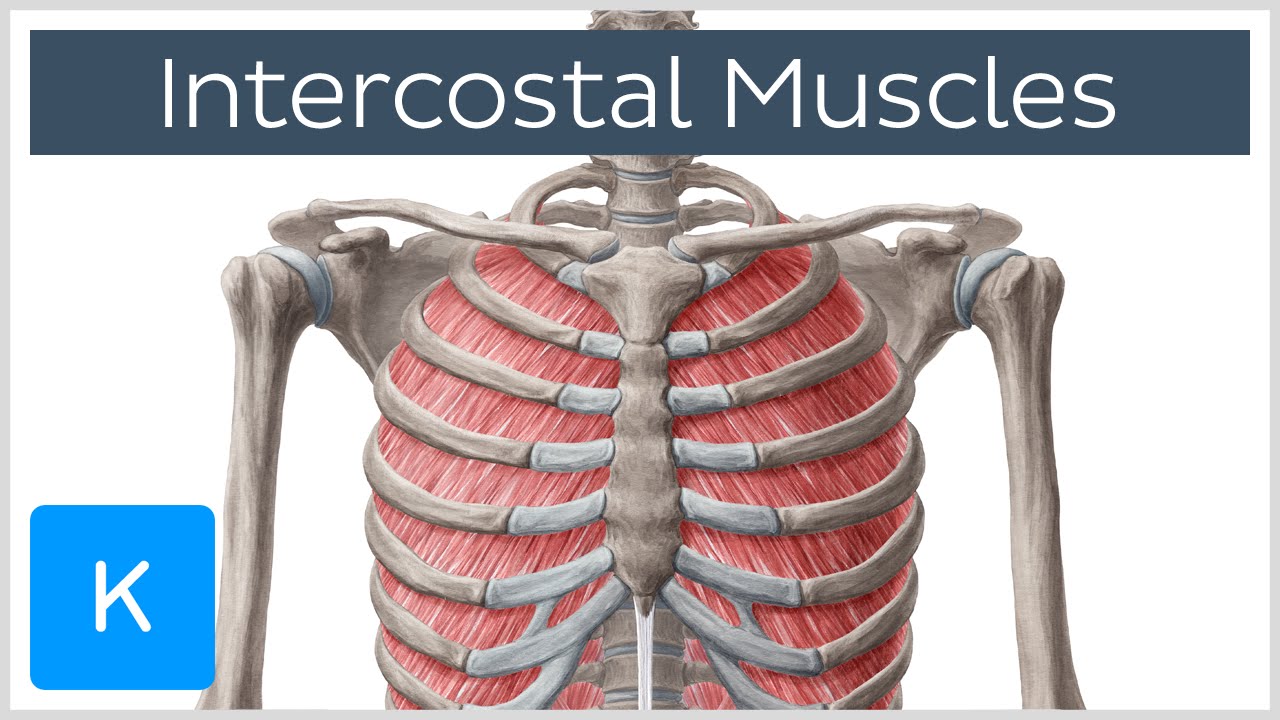
Rib Cage Muscles : Medical Illustration Of Muscular Cage ... from i.ytimg.com In this video we discuss the structure of the rib cage or thoracic cage. Rib cage anatomy the rib cage, shaped in a mild cone shape and more flexible than most bone sets, is made up of varying elements such as the thoracic the top edge of the manubrium has a depression called the suprasternal or jugular notch. Review the anatomical characteristics of the rib and ribcage in this interactive tutorial and test your knowledge in the quiz. Medical human chest skeletal bone structure model. A doctor will diagnose the underlying cause by a physical examination and imaging scans. In humans the rib cage also known as the thoracic cage is a bony and cartilaginous structure which surrounds the thoracic cavity and supports the shoulder girdle to form the core part of the human. The rib cage is one of the strongest structures in the human body, designed to protect two of the most important organ systems: Smartdraw includes 1000s of professional healthcare and anatomy chart templates that you can modify and make your own.
A doctor will diagnose the underlying cause by a physical examination and imaging scans.
Learn vocabulary, terms and more with flashcards, games and other study tools. Rib cage diagram with organs find out more about rib cage diagram with organs. Rib cage anatomy the rib cage shaped in a mild cone shape and more flexible than most bone sets is made up of varying elements such as the thoracic vertebra 12 equally three of those connect to non costal cartilage. Introduction to the structure of the ribcage and ribs: Caring for your ribcage and abdomen sharing the health. The rib cage is the arrangement of ribs attached to the vertebral column and sternum in the thorax of most vertebrates, that encloses and protects the heart. Smartdraw includes 1000s of professional healthcare and anatomy chart templates that you can modify and make your own. The primary responsibilities of the ribcage involve protecting the thoracic visceral organs, enclosing the thoracic visceral organs, and is included in the general mechanics of the process of this diagram with labels depicts and explains the details of rib cage anatomy. Although each rib has its own rom (occurring primarily at the costovertebral joint), rib. Manubrium body xiphoid process sternal angle (of louis). Pain coming from a person's rib cage may be nothing serious, or it may be a medical emergency there are many possible causes of rib cage pain. Moving during chest expansion to enable lung inflation. Create healthcare diagrams like this example called position of lungs in rib cage in minutes with smartdraw.